
A technical, implementation-focused summary of a paired-biopsy study examining the clinical, histologic and immunophenotypic expression of PIILIF across dissecting cellulitis disease states.
Clinical premise
Dissecting cellulitis of the scalp is conventionally classified as a neutrophilic primary cicatricial alopecia characterized by follicular occlusion, rupture, abscess formation, sinus tracts, granulation tissue and scarring.
Those features define active destructive disease. They do not fully account for:
- Relapse after apparent clinical quiescence
- Extension into adjacent scalp
- Reactivation of clinically quiet nodules
- Overlap with other folliculocentric scarring disorders
The study tested whether PIILIF, an upper-follicle lymphocytic-fibrotic pattern previously described in clinically normal-appearing scalp in other cicatricial alopecias, was also present in DCS outside actively suppurative lesions.

Study rationale and cohort
This was a retrospective, exploratory, single-center clinicopathologic study of consecutive patients with clinicopathologically concordant DCS evaluated from December 2022 through November 2024.
The cohort included:
- 12 men
- Mean age of 30 years
- Age range of 21 to 46 years
- Nine patients with an active lesion at lesional biopsy
- Three patients with no active lesions but a clinically inactive residual nodule
- Three patients with concurrent histologic AKN
- One patient with PIILIF identified in a sideburn biopsy
Each patient underwent a protocolized paired-biopsy evaluation during routine care.
Biopsy-site definitions
Clinically normal-appearing scalp, or cNAS
Scalp without visible:
- Nodules
- Pustules
- Sinus tracts
- Alopecia
The area was normal on inspection and palpation at the time of biopsy.
Trichoscopy was used to identify subtle perifollicular abnormalities, including mild erythema, hyperpigmentation or scale, to guide site selection. The cNAS biopsy was typically obtained from parietal scalp approximately 2 to 3 cm from the nearest clinically evident lesion.

Active DCS lesion
A lesion showing one or more of the following:
- Erythema
- Tenderness
- Drainage
- Fluctuance
Clinically inactive, or quiet, nodule
A residual indurated nodule without signs of active inflammation.
Paired-biopsy method
Each patient underwent:
- A 6 mm punch biopsy from cNAS
- A second 6 mm punch biopsy from an active DCS lesion or, when no active lesion was present, a clinically inactive nodule
Both specimens were obtained from the same scalp during the same clinical evaluation.
The paired design allowed comparison of the clinically visible disease state with scalp that appeared unaffected in the same patient.

Sectioning and histopathologic assessment
Each specimen was processed with both:
- Vertical sections
- Transverse sections
Vertical sections were used to assess upper-follicle inflammation, while transverse sections improved visualization of concentric perifollicular fibrosis and adnexal loss.
PIILIF was defined as:
- A perifollicular lymphocytic infiltrate centered on the infundibulum and/or isthmus
- Associated perifollicular lamellar fibroplasia or early concentric fibrosis
- No alternative dominant diagnostic pattern involving the interfollicular epidermis
Two board-certified dermatopathologists independently reviewed the slides while blinded to clinical information, sampling site, lesion activity and treatment history. Differences were resolved by consensus.
Immunohistochemistry
The study used:
- CD4 and CD8 to characterize the relative T-cell population
- CD117 to support mast-cell identification
- Alpha-smooth muscle actin, or alpha-SMA, when fibrosis was equivocal on hematoxylin and eosin staining
Immunohistochemistry was evaluable in eight active-lesion and cNAS pairs.
Across disease states:
- Infiltrates were consistently CD4-predominant
- CD117-positive mast cells were prominent
The findings support an immunologically active upper-follicle process rather than nonspecific end-stage scar alone.
Findings across the three sampled states
Clinical state | PIILIF finding | Suppurative or destructive findings | Sebaceous gland finding |
Normal-appearing scalp | Present in 12 of 12 biopsies | No neutrophilic infiltrates in the nine matched comparisons; no granulation tissue | No complete gland loss in the nine matched comparisons |
Clinically inactive nodules | Present in 3 of 3 nodules | No destructive suppurative features identified | Sebaceous glands preserved |
Active DCS lesions | Infundibulo-isthmic fibrosis in 9 of 9; lymphocytes in 8 of 9 | Neutrophils in 9 of 9; granulation tissue in 5 of 9; follicular destruction or rupture in 6 of 9 | Complete gland loss in 6 of 9 |
Active lesions therefore showed classic neutrophilic destruction and tissue remodeling superimposed on an upper-follicle PIILIF background.
Interpretation
A possible field effect
The presence of PIILIF in cNAS, quiet nodules and active lesions supports a field-effect hypothesis in which low-grade upper-follicle inflammation and early fibrosis extend beyond clinically obvious DCS margins.
This model could help explain relapse, extension and overlap with other folliculocentric disorders.
It remains associative. The study cannot establish whether PIILIF precedes, follows or persists after active DCS lesions.
A possible two-layer inflammatory model
The findings support a working model in which:
- A chronic lymphocytic-fibrotic upper-follicle pattern is present across a wider scalp field
- Episodic neutrophil-dominant activation produces follicular rupture, pus, granulation tissue, gland loss and scarring
This model is hypothesis-generating and was not directly tested mechanistically.
The sebaceous-gland distinction
Sebaceous glands were preserved in cNAS and quiet nodules but completely absent in six of nine active lesions.
This separation raises the hypothesis that sebaceous gland loss may mark a transition toward less reversible disease.
The cross-sectional design cannot prove that sequence. A quiet nodule may represent:
- A treated or spontaneously improved lesion
- An earlier component of a larger lesion
- A biopsy taken outside a destructive focus
Dandruff-related diagnostic discordance
Three patients had previously been managed as having seborrheic dermatitis, but none of the study biopsies supported that diagnosis.
This supports a low threshold for reconsidering the diagnosis when persistent perifollicular erythema or scale does not fit the clinical course.
Prior antifungal or corticosteroid exposure may have attenuated seborrheic dermatitis findings, so the study does not establish that every previous dandruff diagnosis was incorrect.
Overlap with AKN and disease beyond the scalp
Concurrent histologic AKN was present in three of 12 patients. PIILIF was also found in a sideburn specimen from one patient.
These observations support investigation of broader folliculocentric susceptibility in selected patients, but they are too limited to establish prevalence or a universal overlap phenotype.
One-Page Paired-Biopsy Request Guide
Research-informed protocol based on the published study. This is not a formal consensus guideline and should be adapted to the clinical question.
Clinical indication
Consider a paired-biopsy approach when evaluating:
- Recurrent or extending DCS
- Persistent disease despite conventional therapy
- Clinically quiet nodules with a history of reactivation
- Discordance between symptoms and visible findings
- Suspected DCS and AKN overlap
- Persistent perifollicular scale or erythema previously attributed to seborrheic dermatitis
- Suspected extension beyond the visibly affected scalp
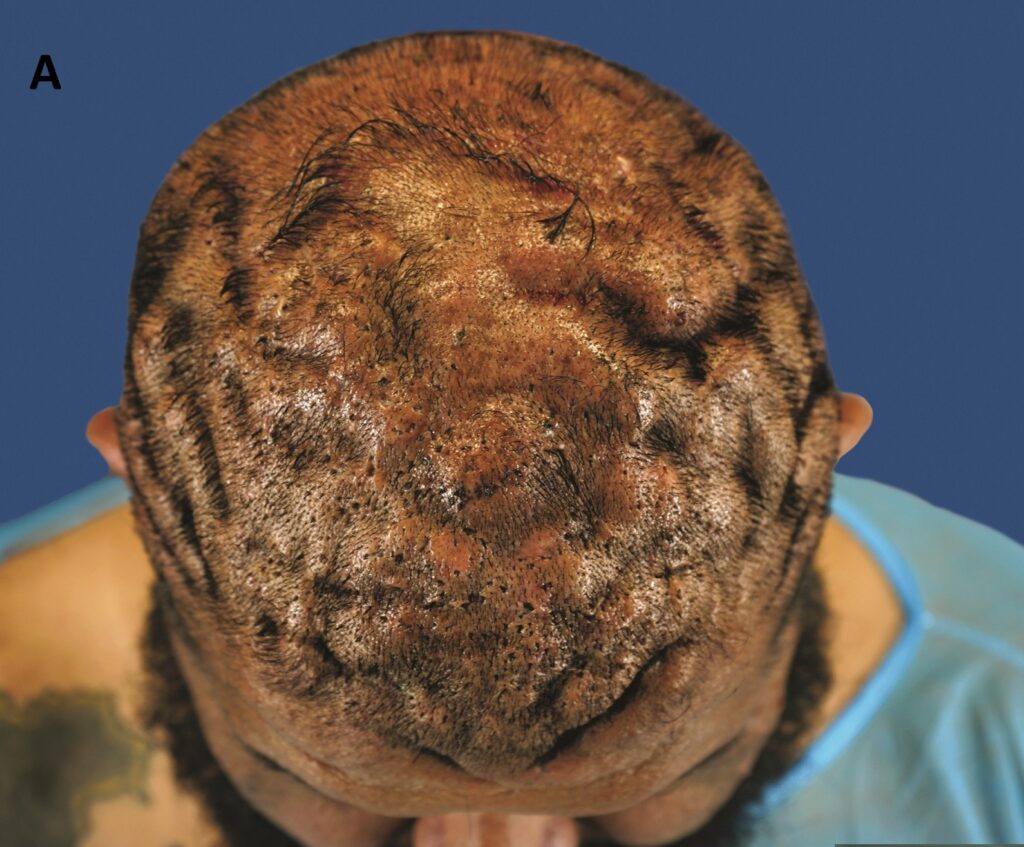
Specimen A: Clinically normal-appearing scalp
Select a site that has:
- No visible nodule
- No pustule
- No sinus tract
- No clinically evident alopecia
- No palpable active lesion
Preferred study location:
- Parietal scalp
- Approximately 2 to 3 cm from the nearest visible lesion
- Guided by subtle trichoscopic perifollicular erythema, hyperpigmentation and/or scale
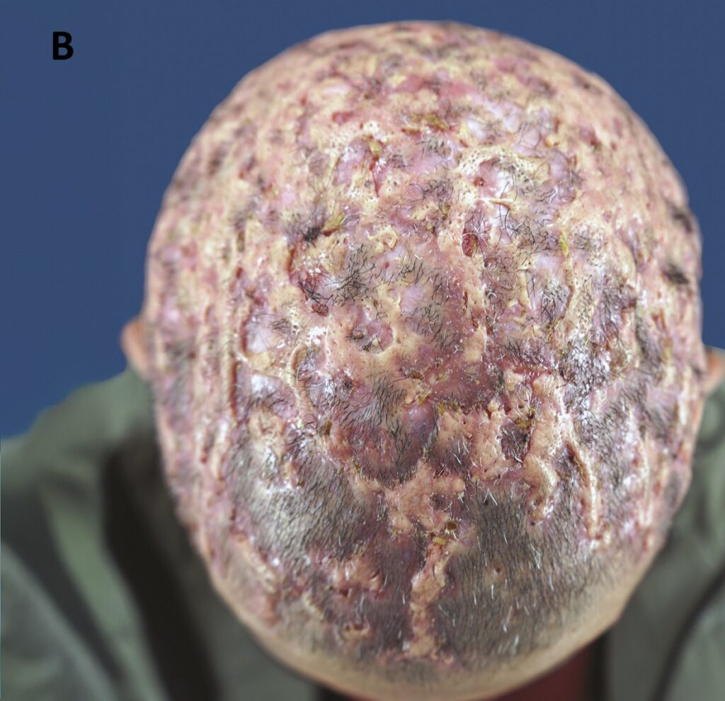
Specimen B: Lesional scalp
When active disease is present, target a lesion showing:
- Erythema
- Tenderness
- Drainage
- Fluctuance
When no active lesion is present, target:
- A residual, firm, clinically inactive nodule
Collection
- Obtain separate 6 mm punch biopsies
- Place specimens in separately labeled containers
- Record exact scalp location
- Label each as cNAS, active lesion or inactive nodule
- Document whether specimens were obtained during the same evaluation
- Include recent and prior treatment exposure
Processing request
Request:
- Vertical sections
- Transverse sections
- Adequate sampling of the upper follicle and surrounding dermis
Histopathologic features to assess
For both specimens:
- Infundibulo-isthmic lymphocytic infiltrate
- Perifollicular lamellar fibroplasia or early concentric fibrosis
- Sebaceous gland preservation or loss
- Epithelial atrophy
- Bulbar inflammation or fibrosis
- Interfollicular epidermal involvement
- Alternative diagnostic patterns
For active lesions:
- Neutrophilic infiltrates
- Follicular rupture
- Extravasated hair shafts
- Granulation tissue
- Avascular scarring
- Sinus tracts
- Adnexal loss
Suggested ancillary stains
When tissue is sufficient and clinically appropriate:
- CD4
- CD8
- CD117
- Alpha-SMA when early fibrosis is equivocal on routine staining
Suggested requisition wording
Clinical history of recurrent or suspected dissecting cellulitis. Paired specimens submitted from clinically normal-appearing scalp and an active lesion or clinically inactive nodule. Please evaluate for perifollicular infundibulo-isthmic lymphocytic inflammation and fibrosis, sebaceous gland status, follicular rupture, neutrophilic inflammation, granulation tissue and scarring. Please process with vertical and transverse sections. Consider alpha-SMA, CD4, CD8 and CD117 if tissue and findings permit.
Limitations
The study’s main limitations include:
- Small sample size
- Single specialty-clinic cohort
- Referral and spectrum bias
- Retrospective design
- Cross-sectional sampling
- No healthy or disease control group
- Heterogeneous treatment exposure before biopsy
- No standardized treatment timing
- Only three inactive nodules
- No treatment-stratified analysis
- Categorical rather than quantitative histologic scoring
- No prespecified interobserver agreement analysis
- No proof of temporal sequence
- No treatment comparison
The 100% cNAS finding should therefore not be interpreted as proof that PIILIF is universal or specific to all DCS cases.
Unanswered research questions
The publication creates several testable questions:
- Does PIILIF in cNAS precede the development of visible DCS lesions?
- Does the burden or distribution of PIILIF predict extension or relapse?
- Does sebaceous gland loss mark a transition toward less reversible disease?
- Are quiet nodules early lesions, partially controlled lesions or post-destructive remnants?
- What molecular event converts a lymphocytic-fibrotic field into a neutrophil-dominant flare?
- How do DHT signaling, cytokine activity, mast cells, microbiome changes and follicular rupture interact?
- Is PIILIF specific to DCS or shared across a broader group of cicatricial alopecias?
- Does addressing PIILIF in addition to suppressing active neutrophilic inflammation improve long-term outcomes?
Prospective longitudinal studies with standardized clinical scores, serial paired biopsies, healthy or disease controls, quantitative pathology and spatial immune analysis are needed. Interventional studies will be required before any treatment-specific conclusions can be made.
Refer a DCS Case
The clinician referral form should request:
- Patient age and sex
- Disease duration
- Current activity status
- Distribution of lesions
- History of drainage, pain, tunnels and scarring
- Prior biopsy reports
- Prior and current treatments
- Scalp photographs
- Trichoscopy images
- History or examination findings of AKN
- History of acne, hidradenitis suppurativa or pilonidal disease
- Suspected facial or beard-area extension
- Reason for referral
- Co-management preference
Request the Paired-Biopsy Protocol
Provide a downloadable one-page PDF version of the protocol above, with:
- Site definitions
- Sampling map
- Specimen labels
- Processing instructions
- Suggested requisition wording
- Dermatopathology checklist
Express Interest in a Multicenter PIILIF Study
The collaboration form should request:
- Clinician name and credentials
- Institution or practice
- City and country
- Estimated annual DCS case volume
- Access to trichoscopy
- Ability to perform paired scalp biopsies
- Dermatopathology support
- Institutional review board capability
- Interest in prospective registry participation
- Interest in serial biopsy studies
- Interest in treatment trials
- Preferred contact information
Publication
Perifollicular Lymphocytic Inflammation and Fibrosis in Dissecting Cellulitis: Evidence of a Consistent Histopathologic Pattern
Published in Clinical, Cosmetic and Investigational Dermatology on July 17, 2026.
Publication-Link Implementation
Link the full paper title on both pages to: https://doi.org/10.2147/CCID.S614816